Introduction to Medical Image Segmentation
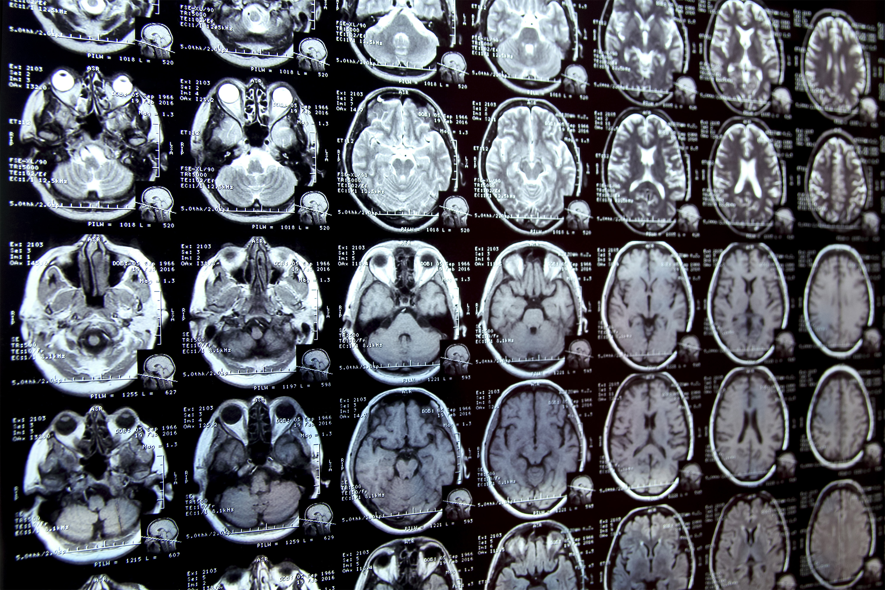
Annotating regions of interest in medical images, a process known as segmentation, is often one of the first steps clinical researchers take when running a new study involving biomedical images. For instance, to determine how the size of the brain’s hippocampus changes as patients age, the scientist first outlines each hippocampus in a series of brain scans. This is often a manual process that can be extremely time-consuming, especially if the regions being studied are challenging to delineate.
The Challenge of Manual Segmentation
Manual image segmentation is so time-consuming that many scientists might only have time to segment a few images per day for their research. This limitation can significantly hinder the pace of medical research and the development of new treatments. The need for an efficient tool to accelerate this process is evident, as it could enable clinical researchers to conduct studies they were previously unable to undertake due to the lack of such a tool.
Developing an AI-Based Solution
To address this challenge, MIT researchers developed an artificial intelligence-based system that enables a researcher to rapidly segment new biomedical imaging datasets by clicking, scribbling, and drawing boxes on the images. This new AI model uses these interactions to predict the segmentation. The system, known as MultiverSeg, combines the benefits of interactive segmentation and task-specific AI models, allowing it to learn from user interactions and improve its predictions over time.
How MultiverSeg Works
MultiverSeg’s architecture is specially designed to use information from images it has already segmented to make new predictions. As the user marks additional images, the number of interactions they need to perform decreases, eventually dropping to zero. The model can then segment each new image accurately without user input. This capability makes MultiverSeg highly efficient and flexible, suitable for a wide range of biomedical imaging applications.
Streamlining Segmentation
Unlike other medical image segmentation models, MultiverSeg allows the user to segment an entire dataset without repeating their work for each image. The interactive tool does not require a presegmented image dataset for training, so users don’t need machine-learning expertise or extensive computational resources. They can use the system for a new segmentation task without retraining the model, making it highly accessible and user-friendly.
Comparison with Existing Tools
When compared to state-of-the-art tools for in-context and interactive image segmentation, MultiverSeg outperformed each baseline. It required less user input with each image, needing only two clicks from the user to generate a segmentation more accurate than a model designed specifically for the task by the ninth new image.
Benefits and Future Directions
The tool’s interactivity enables the user to make corrections to the model’s prediction, iterating until it reaches the desired level of accuracy. This feature, combined with its efficiency, could significantly accelerate studies of new treatment methods and reduce the cost of clinical trials and medical research. Moving forward, the researchers aim to test MultiverSeg in real-world situations with clinical collaborators and improve it based on user feedback, with plans to enable it to segment 3D biomedical images.
Conclusion
MultiverSeg represents a significant advancement in medical image segmentation, offering a powerful, efficient, and user-friendly tool for clinical researchers. By streamlining the segmentation process, it has the potential to unlock new scientific discoveries and improve the efficiency of clinical applications, such as radiation treatment planning. As medical research continues to rely heavily on the analysis of biomedical images, innovations like MultiverSeg will play a crucial role in accelerating progress and saving lives.
FAQs
- What is medical image segmentation?
Medical image segmentation is the process of annotating regions of interest in medical images, which is crucial for various clinical research studies and applications. - How does MultiverSeg improve upon existing segmentation tools?
MultiverSeg combines the benefits of interactive segmentation and task-specific AI models, allowing it to learn from user interactions and improve its predictions over time, requiring less user input and no retraining for new tasks. - What are the potential applications of MultiverSeg?
MultiverSeg can be used to accelerate studies of new treatment methods, reduce the cost of clinical trials, improve the efficiency of clinical applications like radiation treatment planning, and enhance the overall pace of medical research. - Does MultiverSeg require machine-learning expertise or extensive computational resources?
No, MultiverSeg is designed to be accessible and user-friendly, allowing users to apply it to new segmentation tasks without needing machine-learning expertise or retraining the model. - What are the future plans for MultiverSeg?
The researchers plan to test MultiverSeg in real-world clinical settings, gather user feedback for improvement, and develop its capability to segment 3D biomedical images.


